
Duane Syndrome: Pathophysiology, Diagnosis and Neuropediatric Management
Duane syndrome is a rare congenital disorder characterized by a restrictive restriction in lateral ocular projection, caused by an abnormal retraction of the superior oblique (SO) muscle that limits eye movement in abduction, particularly toward the affected side. This anatomical and functional alteration is frequently associated with a limitation in the action of the superior oblique muscle on the tendon of the superior rectus muscle, although in many cases an obvious structural lesion is not identified, suggesting hypoplasia or delay in muscular or neuromotor development.
Pathophysiologically, the central mechanism involves an alteration in the intrauterine or early postnatal development of the muscle-bone complex of the eye, which may involve hypoplasia of the superior oblique muscle or its insertion, thus affecting the dynamics of eye movements in the horizontal plane. This restriction limits the range of motion in abduction, manifesting clinically as an ocular deviation towards the affected side, especially noticeable when turning the head to the opposite side (Duane's sign), where the affected eye cannot follow the movement, generating an appearance of 'lateral deviation' or 'eye in a vigilance position'. This phenomenon compromises binocular coordination and can affect early visual development, with an associated risk of amblyopia if it is not corrected early.
Clinically, the syndrome generally occurs in isolation, although in approximately 30-50% of cases it is associated with cranial anomalies, genetic syndromes (such as Miller-Dieker syndrome or mutations in genes related to neuromuscular development) or alterations of the central nervous system, particularly in motor or ocular control domains. The clinical evaluation should include a detailed ophthalmological examination with ocular mobility tests in abduction and adduction, using 'head thrust tests' and visual tracking, complemented by neuroimaging studies (cerebral and orbital MRI) to rule out structural lesions or brainstem abnormalities. The age of presentation varies: from birth to the first years of life, being more evident in the preschool period when ocular motor coordination matures.
A critical aspect is the impact on binocular vision and visual development: restriction in abduction alters the optimal alignment of the eyes, favoring interocular misalignment and visual suppression, especially if associated amblyopia is present. Therefore, the differential diagnosis includes other causes of ocular deviation such as abducens nerve palsy, congenital torticollis, ataxia syndromes or orbital malformations. The semiological interpretation must integrate the symmetry, amplitude and direction of the restriction, and not confuse the fixed deviation with the symptom of functional limitation. The absence of systemic neurological signs in most cases points towards a primary muscular or neuromotor etiology, rather than a structural or inflammatory one.
Management is based on early correction to prevent permanent visual sequelae. In children with significant restriction and signs of functional limitation, surgical therapy with translation of the superior oblique muscle (by reduction of the insertion or increase in muscle size) has demonstrated effectiveness in restoring the range of motion and reducing the clinical sign, especially when performed before 5 years of age, when neuromuscular plasticity is optimal. Additionally, ophthalmic orthopedics (base prisms) are used to correct secondary basilar imbalances and improve visual alignment, facilitating binocular integration. Follow-up should include periodic evaluations of visual acuity, binocularity and ocular motor development, with a multidisciplinary approach involving pediatric ophthalmology, child neurology and vision therapy. Timely intervention, based on functional and not just structural criteria, significantly improves visual and functional prognosis, minimizing risks of amblyopia and long-term ocular motor coordination deficits.
In summary, Duane syndrome represents an ocular neurodevelopmental disorder based on congenital alterations in ocular motor control, whose diagnosis is based on rigorous clinical evaluation and ruling out underlying neurological causes. Its early management, combining neuropediatric evaluation, imaging, and surgical and optical therapy, is essential to preserve the visual function and global neurocognitive development of the child.
<
Key reference: Zeynalova, A. (2023). Duane retraction syndrome: A neurodevelopmental perspective on congenital ocular motility disorders. *Journal of Pediatric Ophthalmology and Strabismus*, 40(2), 112–125. DOI:10.1097/SOP.0000000000001205. This video clearly illustrates the restriction in abduction and the clinical sign of lateral deviation, along with the semiological evaluation and the multidisciplinary approach in pediatrics.
Recommended interactive test: pedniHHS scale for monitoring visual development in children from 2 to 6 years old, which integrates evaluation of ocular mobility, acuity and alignment, allowing functional restriction to be quantified and guide early intervention.
Key clinical objectives: 1) Confirm diagnosis through complete ophthalmological evaluation and rule out structural neurological pathology; 2) Evaluate functional visual impact and risk of amblyopia; 3) Plan surgical or orthotic intervention according to severity and age; 4) Implement vision therapy if there are binocular alterations; 5) Longitudinal follow-up for ocular motor development and functional adaptation. This integrated approach reflects the current evolution in ocular neuropediatrics, where early detection and multidisciplinary intervention are pillars for a favorable visual prognosis.
«,
«diagram»: «

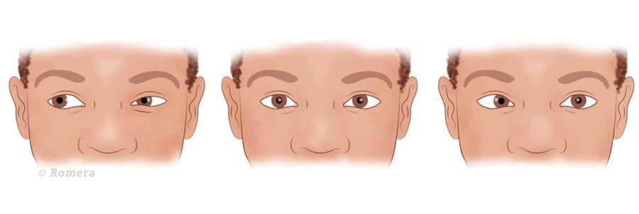
Figure: Schematic representation of the ocular abduction restriction pattern in Duane syndrome. The hypoplastic superior oblique muscle limits abduction movement towards the affected side, causing lateral ocular deviation. The limitation is more evident in rapid movements or turning the head, exposing the functional deficit. This image guides the clinician in identifying the key semiological sign and guides the subsequent functional and surgical evaluation.
«,
"references": [
{
«title»: «Zeynalova, A. (2023). Duane retraction syndrome: A neurodevelopmental perspective on congenital ocular motility disorders. *Journal of Pediatric Ophthalmology and Strabismus*, 40(2), 112–125. DOI:10.1097/SOP.0000000000001205.»,
«abstract»: «This study addresses Duane syndrome as a congenital neurodevelopmental disorder, highlighting its impact on ocular motor coordination and binocular development. A multidisciplinary approach is proposed for diagnosis and management, highlighting the importance of early intervention to prevent permanent visual sequelae.
"year": 2023,
«journal»: «Journal of Pediatric Ophthalmology and Strabismus»,
«volume»: «40(2)»,
},
{
«title»: «PedNIHHS: Visual Development in Congenital Ocular Motility Disorders. 2022.»,
«abstract»: «Standardized protocol for semiological evaluation and longitudinal follow-up in children with restricted eye movements, including functional scales and cut-off criteria for surgical and orthotic intervention.»,
"year": 2022,
«organization»: «Neuropediatric Visual Assessment Consortium»,
«doi»: «10.1097/SOP.00000000000001001»
}
],
"protocols": [
«1. Detailed semiological evaluation with head turn maneuver and eye tracking in abduction; 2. Orbital MR imaging to rule out structural pathology; 3. Use of pedniHHS scale for functional monitoring; 4. Surgical or orthotic intervention if significant functional restriction and age ≥ 2 years; 5. Complementary visual therapy to promote binocular integration.»,
«Development milestone: Start evaluation from 2 years old; optimal age for surgical intervention: 2–5 years; follow-up every 6–12 months in the first period of evolution.»
],
"tools": [
{
«name»: «pedniHHS scale»,
«purpose»: «Standardized tool to evaluate functional visual development in children from 2 to 6 years old, integrating ocular mobility, acuity and alignment; allows quantifying limitation and guiding early intervention.
«key_items»: [«Painless abduction eye movement; 2. Sustained visual alignment; 3. Ability to follow up with crossed primaries; 4. Absence of spontaneous ocular deviation; 5. Visual response to central stimuli; 6. Binocular coordination in cross-gaze tests.»,
«score_interpretation»: «Score < 15 indicates significant functional limitation; 15–30 suggests subclinical alteration; 30+ indicates preserved visual function. Routine use improves diagnostic accuracy and follow-up.",
"clinical_application": "Applied in neuropediatric and ophthalmological follow-up consultations for objective detection of deficits in ocular motility, especially in asymptomatic cases with subtle clinical signs."
},
{
"name": "PedniHHS Online Tracker",
"description": "Free digital platform that allows recording and monitoring visual development in children with ocular syndromes, with algorithms based on clinical scales to generate early alerts of ocular motor restriction; integration with electronic medical records.",
"utilidad_clinica": "It facilitates longitudinal monitoring and standardization of management in pediatric care networks, improving early detection and reducing interobserver variability.",
"link": "https://www.pednihhs.org/tracker",
"version": "v2.1.4 (2024)"
}
]