
In the realm of neuroscience, electroencephalography (EEG) stands as a pivotal tool for unraveling the mysteries of the brain. It allows researchers and clinicians to observe the electrical activity generated by the brain’s neurons, offering insights into various neurological conditions. Among the myriad of waveforms discernible in an EEG, one intriguing pattern is that of wicket spikes. These peculiar formations hold significance in understanding brain function, yet they can also lead to misinterpretations, particularly when juxtaposed with epileptiform activity.
What are Wicket Spikes?
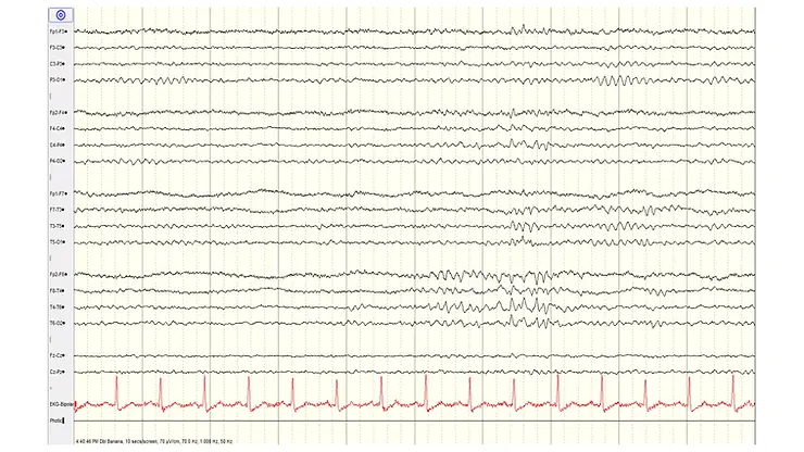
Wicket spikes, also known as wicket rhythms or wicket waves, are distinctive benign EEG patterns characterized by their unique appearance resembling the silhouette of a cricket wicket, hence the name. These patterns typically manifest as sharply contoured, repetitive waveforms with a «sawtooth» or «spiky» appearance. Wicket spikes are often observed in the central and temporal regions of the brain, although they can occur elsewhere.
Interpreting Wicket Spikes in EEG:
Wicket spikes are typically considered benign and non-epileptiform in nature. They are often associated with various physiological and non-pathological states, including drowsiness, sleep, or relaxation. In such contexts, wicket spikes may arise due to the modulation of neuronal firing patterns by sleep-related neurotransmitters or changes in arousal levels.
Furthermore, wicket spikes can also emerge during hyperventilation or in response to sensory stimuli, reflecting the brain’s dynamic adaptability to environmental cues. In these instances, wicket spikes serve as indicators of normal physiological processes rather than pathological conditions.
Misinterpretations and Epileptiform Activity:
Despite their benign nature, wicket spikes can sometimes be mistaken for epileptiform activity, particularly by inexperienced or untrained observers. Epileptiform discharges, such as spikes, sharp waves, or spike-and-wave complexes, share certain visual similarities with wicket spikes, thereby posing challenges in accurate EEG interpretation.
This potential for misinterpretation underscores the importance of comprehensive clinical evaluation and contextual analysis when interpreting EEG findings. Factors such as the patient’s clinical history, concurrent symptoms, and other EEG abnormalities must be carefully considered to differentiate between wicket spikes and epileptiform activity accurately.

Distinguishing Features:
Several distinguishing features can aid in the differentiation between wicket spikes and epileptiform discharges:
- Temporal Dynamics: Wicket spikes typically exhibit a rhythmic, repetitive pattern over time, often occurring in clusters or runs. In contrast, epileptiform discharges may demonstrate greater variability in morphology, duration, and inter-event intervals.
- Topographic Distribution: Wicket spikes commonly localize to specific cortical regions, such as the central or temporal regions, and often display bilateral symmetry. In contrast, epileptiform discharges may exhibit more variable topographic distributions, depending on the underlying epileptogenic focus.
- Associated Clinical Context: Assessing the clinical context surrounding EEG recordings is crucial for accurate interpretation. Wicket spikes occurring during periods of relaxation, drowsiness, or sensory stimulation are more likely to be benign, whereas epileptiform discharges may coincide with seizure activity or neurological symptoms.
Frequency and Patient Population:
Wicket spikes typically occur in the frequency range of 6 to 11 Hz, reflecting their association with states of reduced arousal or vigilance, such as drowsiness or relaxation. They are commonly observed in adults over the age of 30 during various stages of sleep, including light sleep and drowsiness. Additionally, wicket spikes may also manifest during states of relaxation or during specific EEG protocols such as hyperventilation.
Conclusion:
In conclusion, wicket spikes represent a fascinating EEG phenomenon with distinctive characteristics and physiological correlates. While generally benign, their resemblance to epileptiform activity underscores the importance of cautious interpretation and integration with clinical context. By understanding the unique features of wicket spikes and their potential pitfalls, clinicians can navigate EEG interpretations more effectively, enhancing diagnostic accuracy and patient care.
Wicked spikes.
En el campo de la neurociencia, la electroencefalografía (EEG) se presenta como una herramienta fundamental para desentrañar los misterios del cerebro. Permite a investigadores y clínicos observar la actividad eléctrica generada por las neuronas cerebrales, ofreciendo información sobre diversas condiciones neurológicas. Entre la gran variedad de formas de onda discernibles en un EEG, un patrón intrigante es el de los picos wicket. Estas formaciones peculiares tienen importancia para comprender la función cerebral, aunque también pueden conducir a interpretaciones erróneas, particularmente cuando se comparan con actividad epileptiforme. ¿Qué son los Picos Wicket? Los picos wicket, también conocidos como ritmos wicket u ondas wicket, son patrones de EEG benignos distintivos caracterizados por su apariencia única que se asemeja a la silueta de un wicket de críquet, de ahí el nombre. Estos patrones típicamente se manifiestan como formas de onda repetitivas de contorno agudo con una apariencia de «diente de sierra» o «puntiaguda». Los picos wicket se observan frecuentemente en las regiones centrales y temporales del cerebro, aunque pueden ocurrir en otros lugares. Interpretación de los Picos Wicket en el EEG: Los picos wicket generalmente se consideran benignos y de naturaleza no epileptiforme. A menudo se asocian con varios estados fisiológicos y no patológicos, incluyendo somnolencia, sueño o relajación. En tales contextos, los picos wicket pueden surgir debido a la modulación de los patrones de disparo neuronal por neurotransmisores relacionados con el sueño o cambios en los niveles de alerta. Además, los picos wicket también pueden emerger durante la hiperventilación o en respuesta a estímulos sensoriales, reflejando la adaptabilidad dinámica del cerebro a las señales ambientales. En estos casos, los picos wicket sirven como indicadores de procesos fisiológicos normales en lugar de condiciones patológicas. Interpretaciones Erróneas y Actividad Epileptiforme: A pesar de su naturaleza benigna, los picos wicket a veces pueden confundirse con actividad epileptiforme, particularmente por observadores inexpertos o sin entrenamiento. Las descargas epileptiformes, como puntas, ondas agudas o complejos punta-onda, comparten ciertas similitudes visuales con los picos wicket, planteando así desafíos en la interpretación precisa del EEG. Este potencial de interpretación errónea subraya la importancia de una evaluación clínica integral y un análisis contextual al interpretar los hallazgos del EEG. Factores como la historia clínica del paciente, síntomas concurrentes y otras anomalías del EEG deben considerarse cuidadosamente para diferenciar con precisión entre picos wicket y actividad epileptiforme. Características Distintivas: Varias características distintivas pueden ayudar en la diferenciación entre picos wicket y descargas epileptiformes: Frecuencia y Población de Pacientes: Los picos wicket típicamente ocurren en el rango de frecuencia de 6 a 11 Hz, reflejando su asociación con estados de alerta o vigilancia reducida, como somnolencia o relajación. Se observan comúnmente en adultos mayores de 30 años durante varias etapas del sueño, incluyendo sueño ligero y somnolencia. Además, los picos wicket también pueden manifestarse durante estados de relajación o durante protocolos específicos de EEG como la hiperventilación. Conclusión: En conclusión, los picos wicket representan un fenómeno fascinante del EEG con características distintivas y correlatos fisiológicos. Aunque generalmente son benignos, su semejanza con la actividad epileptiforme subraya la importancia de una interpretación cautelosa e integración con el contexto clínico. Al comprender las características únicas de los picos wicket y sus posibles dificultades, los clínicos pueden navegar las interpretaciones del EEG de manera más efectiva, mejorando la precisión diagnóstica y la atención al paciente.
References:
- Niedermeyer, E., & da Silva, F. L. (2005). Electroencephalography: Basic Principles, Clinical Applications, and Related Fields (5th ed.). Lippincott Williams & Wilkins.
- Husain, A. M. (2016). Wicket spikes. In The Clinical Neurophysiology Primer (pp. 201-204). Humana Press, Cham.
- Tatum, W. O. (2013). Artifact and normal variants. In Handbook of EEG Interpretation (pp. 135-162). Demos Medical Publishing.
- Blume, W. T., Kaibara, M., & Specchio, N. (2006). Definition and diagnostic criteria of epileptic seizures and epilepsies: Neurophysiological, EEG, and neuroimaging studies. Epileptic Disorders, 8(S1), S3-S12.
- Pramod Krishnan, Dr. «Non-Epileptiform Variants in EEG.» SlideShare, https://www.slideshare.net/drpramodkrishnan/non-epileptiform-variants-in-eegpptx.