

Comparison of myelin: CNS vs. SNP
Myelin is a lipid envelope that insulates axons to speed up nerve conduction. Its structure and producing cells vary between CNS and PNS.
Origin and composition
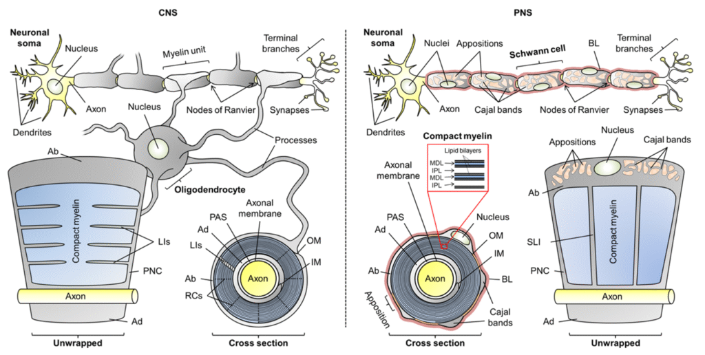
- CNS: Production by oligodendrocytes. A single cell can myelinate up to 50 axons.
- SNP: Production by Schwann cells. Each cell envelops a single axonal segment (~1 mm).
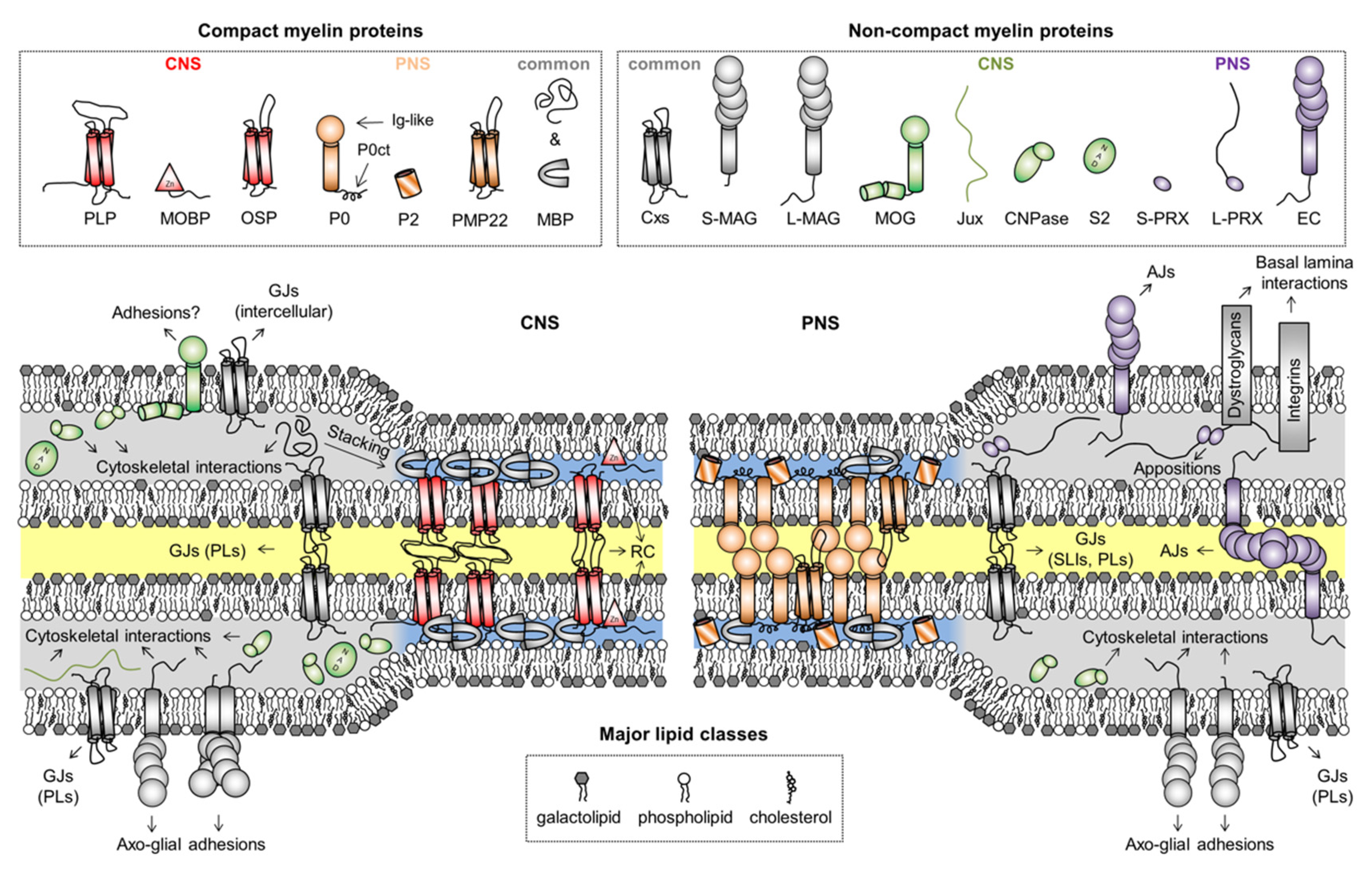
- Key proteins:
- CNS: PLP, MBP (main structural).
- SNP: P0 (adhesion glycoprotein).
Function and vulnerability
- Conduction speed: up to 120 m/s in CNS, slower in PNS.
- Myelin in the CNS is more resistant to inflammation than in the PNS ([1]).
- The PNS is more vulnerable to damage from environmental and inflammatory factors.
Clinical implications
- Multiple Sclerosis (MS): Deterioration of myelin in the CNS due to loss of oligodendrocytes.
- Diagnostic tests:
- TCN and VEP: evaluate myelin function in SNP.
- Magnetic resonance imaging (MRI) and spinal puncture: detect myelinated lesions in the CNS.
- Bell's facial palsy: Damage to peripheral myelin; alterations in TCN and facial evoked potentials ([4]).
Clue: Myelin is essential for rapid nerve transmission. Its evaluation is essential in disorders such as MS and peripheral neuropathies.