
Among the multiple patterns that we can find in EEG waves, one of the most intriguing is wicked spikes. These peculiar formations are important when it comes to understanding brain functioning, but they can also lead to misinterpretation, particularly when they are confused with juxtaposed epileptiform activity.
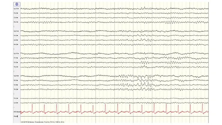
Misinterpretation of wicket spikes.
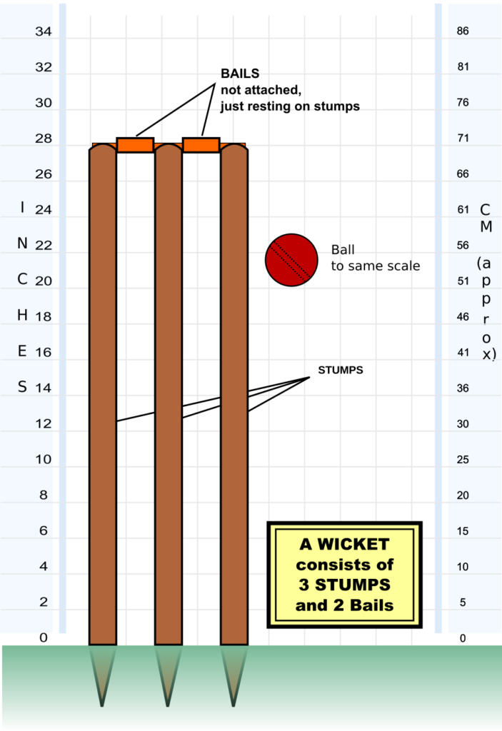
Wicked spikes, also known as wicked rhythms or wicked waves, are a characteristic benign EEG pattern that is identified by a silhouette reminiscent of a "cricket wicket" or cricket target.
This pattern usually manifests itself as a repetitive and sharp wave, with a saw-shaped or dotted shape. They are usually observed in the central and temporal region of the brain, although they can occur in other locations.
They are considered benign and non-epileptiform. They are associated with various physiological states, such as drowsiness, sleep or relaxation. In these contexts, wicket spikes may emerge due to modulation of neuronal firing patterns by sleep-related neurotransmitters.
Furthermore, wicket spikes can emerge during hyperventilation or in response to sensory stimuli, reflecting the brain's ability to adapt to different environmental signals. In these circumstances, the presence of wicket spikes indicates normal brain functioning.
Misinterpretation of wicket spikes.
Despite their benign nature, it is not uncommon for them to be misinterpreted and misidentified as epileptiform activity. Epileptiform discharges, such as spikes, sharp waves, or spike-wave complexes, share certain visual similarities with wicket spikes.

There are several differentiating characteristics:
- Temporal dynamics: Wicket spikes typically present a repetitive rhythmic pattern over time, occurring in clusters. In contrast, epileptiform discharges are more variable, have more heterogeneous inter-event intervals, can have different durations and change morphology from one complex to another.
- Topographic distribution: Wicket spikes are predominantly located in the central and temporal regions, and are usually bilaterally symmetrical.
- Morphology: They also tend to have a harmonious morphology of growth and decrease, "wax and wane." They usually have a frequency between 6 and 11 Hz.
- Associated clinical context: The clinical context is essential for interpretation. It is necessary to know the psychophysiological state during the recording, since these events occur during periods of relaxation, drowsiness or certain sensory stimuli.
References:
- Niedermeyer, E., & da Silva, F. L. (2005). Electroencephalography: Basic Principles, Clinical Applications, and Related Fields (5th ed.). Lippincott Williams & Wilkins.
- Husain, A. M. (2016). Wicket spikes. In The Clinical Neurophysiology Primer (pp. 201-204). Humana Press, Cham.
- Tatum, W. O. (2013). Artifact and normal variants. In Handbook of EEG Interpretation (pp. 135-162). Demos Medical Publishing.
- Blume, W. T., Kaibara, M., & Specchio, N. (2006). Definition and diagnostic criteria of epileptic seizures and epilepsies: Neurophysiological, EEG, and neuroimaging studies. Epileptic Disorders, 8(S1), S3-S12.
- Pramod Krishnan, Dr. “Non-Epileptiform Variants in EEG.” SlideShare, https://www.slideshare.net/drpramodkrishnan/non-epileptiform-variants-in-eegpptx.