
Timing for performing MRI studies: Clinical and Pathophysiological Considerations
Depending on age, there are different peculiarities and limitations:
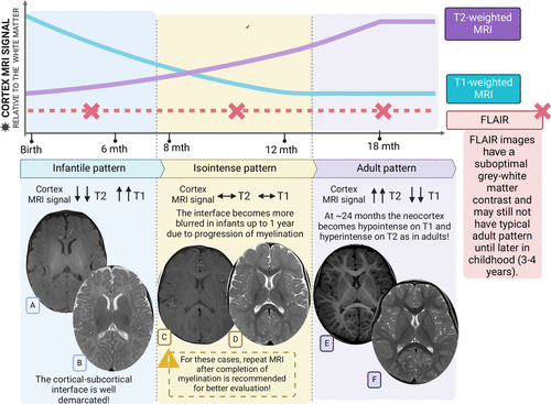
Performing magnetic resonance imaging (MRI) in children is crucial for diagnosing various neurological conditions. However, it is important to consider the patient's age, as tissue immaturity during childhood may lead to incorrect or insufficient interpretations. The justification for repeating MRI studies varies significantly depending on age. For patients under 6 months of age, MRI may be inconclusive due to immaturity of gray and white matter tissues, justifying repeat imaging between 6 and 12 months for improved resolution. In children aged 6 to 12 months, the gray-to-white matter ratio improves, allowing better interpretation, but repeat imaging is necessary if unclear suspicions remain. For ages 12 to 24 months, MRI is ideal for characterizing dysplasias or abnormal migrations. Finally, in patients over 2 years of age, MRI can be definitive in many cases, although repetition is only warranted if there are clinical changes.
Depending on the clinical indication and the time of performance, it may be necessary to repeat:
Repeat MRI depends on the clinical situation and initial indication. In patients with refractory epilepsy, an initial MRI is recommended between 1 and 2 years of age, and repeat imaging should be performed if disease onset was early. For global developmental delay, an initial MRI is necessary between 6 and 24 months of age, and repeat imaging should be done if symptoms progress or new ones emerge. In cases of suspected prenatal congenital malformations of cortical development (MCD), immediate postnatal MRI is recommended, with a follow-up scan at 6 to 12 months for improved resolution. In symptomatic neonates, initial MRI should be performed before 6 months (if urgent) and repeated between 6 and 12 months to confirm findings. For fetal MRI, an initial study is required if schizencephaly or lissencephaly was suspected, with repeat imaging during the first year of life to adequately characterize abnormalities.
| Clinical situation | Initial MRI | Control MRI |
|---|---|---|
| Refractory epilepsy | 1–2 years | Repeat if it was early or normal initial |
| Global developmental delay | 6–24 months | If symptoms progress or new ones arise |
| Prenatal suspicion of DCM | Immediate postnatal | At 6–12 months for greater resolution |
| Symptomatic neonates | <6 months (if urgent) | Repeat after 6–12 months to confirm findings |
| fetal MRI | If there was suspicion of schisencephaly or lissencephaly. | Repeat in the first year of life to adequately characterize. |
Moreover, the accuracy in interpreting MRI studies depends on a clear understanding of cerebral physiology and pathology. The immaturity of gray and white matter tissues during childhood may lead to misinterpretations. For example, decreased brain fat in patients with microcephaly may be incorrectly interpreted as hypoglycemia without proper image interpretation. Cortical development malformations (MCD) can exhibit variability in the appearance of signs and symptoms, justifying repeat imaging to confirm or clarify initial findings.
19955111 {19955111:MKR777X4} 1 vancouver 50 default 7842 https://neuropediatoolkit.org/wp-content/plugins/zotpress/%7B%22status%22%3A%22success%22%2C%22updateneeded%22%3Afalse%2C%22instance%22%3Afalse%2C%22meta%22%3A%7B%22request_last%22%3A0%2C%22request_next%22%3A0%2C%22used_cache%22%3Atrue%7D%2C%22data%22%3A%5B%7B%22key%22%3A%22MKR777X4%22%2C%22library%22%3A%7B%22id%22%3A19955111%7D%2C%22meta%22%3A%7B%22creatorSummary%22%3A%22Brunelli%20et%20al.%22%2C%22parsedDate%22%3A%222024-11%22%2C%22numChildren%22%3A1%7D%2C%22bib%22%3A%22%26lt%3Bdiv%20class%3D%26quot%3Bcsl-bib-body%26quot%3B%20style%3D%26quot%3Bline-height%3A%201.35%3B%20%26quot%3B%26gt%3B%5Cn%20%20%26lt%3Bdiv%20class%3D%26quot%3Bcsl-entry%26quot%3B%20style%3D%26quot%3Bclear%3A%20left%3B%20%26quot%3B%26gt%3B%5Cn%20%20%20%20%26lt%3Bdiv%20class%3D%26quot%3Bcsl-left-margin%26quot%3B%20style%3D%26quot%3Bfloat%3A%20left%3B%20padding-right%3A%200.5em%3B%20text-align%3A%20right%3B%20width%3A%201em%3B%26quot%3B%26gt%3B1.%26lt%3B%5C%2Fdiv%26gt%3B%26lt%3Bdiv%20class%3D%26quot%3Bcsl-right-inline%26quot%3B%20style%3D%26quot%3Bmargin%3A%200%20.4em%200%201.5em%3B%26quot%3B%26gt%3BBrunelli%20JM%2C%20Lopes%20TJP%2C%20Alves%20IS%2C%20Delgado%20DS%2C%20Lee%20HW%2C%20Martin%20MGM%2C%20et%20al.%20Malformations%20of%20Cortical%20Development%3A%20Updated%20Imaging%26%23xA0%3B%20%26%23xA0%3B%20%26%23xA0%3B%20%26%23xA0%3B%20%26%23xA0%3B%20%26%23xA0%3B%20%26%23xA0%3B%20%26%23xA0%3B%20%26%23xA0%3B%20%26%23xA0%3B%20%20Review.%20RadioGraphics%20%5BInternet%5D.%202024%20Nov%20%5Bcited%202025%20May%201%5D%3B44%2811%29%3Ae230239.%20Available%20from%3A%20%26lt%3Ba%20class%3D%26%23039%3Bzp-ItemURL%26%23039%3B%20href%3D%26%23039%3Bhttps%3A%5C%2F%5C%2Fpubs.rsna.org%5C%2Fdoi%5C%2Fabs%5C%2F10.1148%5C%2Frg.230239%26%23039%3B%26gt%3Bhttps%3A%5C%2F%5C%2Fpubs.rsna.org%5C%2Fdoi%5C%2Fabs%5C%2F10.1148%5C%2Frg.230239%26lt%3B%5C%2Fa%26gt%3B%26lt%3B%5C%2Fdiv%26gt%3B%5Cn%20%20%26lt%3B%5C%2Fdiv%26gt%3B%5Cn%26lt%3B%5C%2Fdiv%26gt%3B%22%2C%22data%22%3A%7B%22itemType%22%3A%22journalArticle%22%2C%22title%22%3A%22Malformations%20of%20Cortical%20Development%3A%20Updated%20Imaging%20%20%20%20%20%20%20%20%20%20%20%20%20%20%20%20%20%20%20%20%20Review%22%2C%22creators%22%3A%5B%7B%22creatorType%22%3A%22author%22%2C%22firstName%22%3A%22Julia%20%20%20%20%20%20%20%20%20%20%20%20%20%20%20%20%20%20%20%20%20%20%20%20%20%20%20%20%20M.%22%2C%22lastName%22%3A%22Brunelli%22%7D%2C%7B%22creatorType%22%3A%22author%22%2C%22firstName%22%3A%22Thiago%20J.%20%20%20%20%20%20%20%20%20%20%20%20%20%20%20%20%20%20%20%20%20%20%20%20%20%20%20%20%20P.%22%2C%22lastName%22%3A%22Lopes%22%7D%2C%7B%22creatorType%22%3A%22author%22%2C%22firstName%22%3A%22Isabela%20%20%20%20%20%20%20%20%20%20%20%20%20%20%20%20%20%20%20%20%20%20%20%20%20%20%20%20%20S.%22%2C%22lastName%22%3A%22Alves%22%7D%2C%7B%22creatorType%22%3A%22author%22%2C%22firstName%22%3A%22Daniel%20%20%20%20%20%20%20%20%20%20%20%20%20%20%20%20%20%20%20%20%20%20%20%20%20%20%20%20%20S.%22%2C%22lastName%22%3A%22Delgado%22%7D%2C%7B%22creatorType%22%3A%22author%22%2C%22firstName%22%3A%22Hae%20%20%20%20%20%20%20%20%20%20%20%20%20%20%20%20%20%20%20%20%20%20%20%20%20W.%22%2C%22lastName%22%3A%22Lee%22%7D%2C%7B%22creatorType%22%3A%22author%22%2C%22firstName%22%3A%22Maria%20G.%20%20%20%20%20%20%20%20%20%20%20%20%20%20%20%20%20%20%20%20%20%20%20%20%20%20%20%20%20M.%22%2C%22lastName%22%3A%22Martin%22%7D%2C%7B%22creatorType%22%3A%22author%22%2C%22firstName%22%3A%22Marcos%20F.%20%20%20%20%20%20%20%20%20%20%20%20%20%20%20%20%20%20%20%20%20%20%20%20%20%20%20%20%20L.%22%2C%22lastName%22%3A%22Docema%22%7D%2C%7B%22creatorType%22%3A%22author%22%2C%22firstName%22%3A%22Samya%20%20%20%20%20%20%20%20%20%20%20%20%20%20%20%20%20%20%20%20%20%20%20%20%20S.%22%2C%22lastName%22%3A%22Alves%22%7D%2C%7B%22creatorType%22%3A%22author%22%2C%22firstName%22%3A%22Paula%20%20%20%20%20%20%20%20%20%20%20%20%20%20%20%20%20%20%20%20%20%20%20%20%20C.%22%2C%22lastName%22%3A%22Pinho%22%7D%2C%7B%22creatorType%22%3A%22author%22%2C%22firstName%22%3A%22Vinicius%20%20%20%20%20%20%20%20%20%20%20%20%20%20%20%20%20%20%20%20%20%20%20%20%20%20%20%20%20T.%22%2C%22lastName%22%3A%22Gon%5Cu00e7alves%22%7D%2C%7B%22creatorType%22%3A%22author%22%2C%22firstName%22%3A%22Lucas%20R.%20L.%20%20%20%20%20%20%20%20%20%20%20%20%20%20%20%20%20%20%20%20%20%20%20%20%20%20%20%20%20B.%22%2C%22lastName%22%3A%22Oliveira%22%7D%2C%7B%22creatorType%22%3A%22author%22%2C%22firstName%22%3A%22Jorge%20%20%20%20%20%20%20%20%20%20%20%20%20%20%20%20%20%20%20%20%20%20%20%20%20%20%20%20%20T.%22%2C%22lastName%22%3A%22Takahashi%22%7D%2C%7B%22creatorType%22%3A%22author%22%2C%22firstName%22%3A%22Pejman%20%20%20%20%20%20%20%20%20%20%20%20%20%20%20%20%20%20%20%20%20%20%20%20%20%20%20%20%20J.%22%2C%22lastName%22%3A%22Maralani%22%7D%2C%7B%22creatorType%22%3A%22author%22%2C%22firstName%22%3A%22Camila%20%20%20%20%20%20%20%20%20%20%20%20%20%20%20%20%20%20%20%20%20%20%20%20%20%20%20%20%20T.%22%2C%22lastName%22%3A%22Amancio%22%7D%2C%7B%22creatorType%22%3A%22author%22%2C%22firstName%22%3A%22Claudia%20%20%20%20%20%20%20%20%20%20%20%20%20%20%20%20%20%20%20%20%20%20%20%20%20%20%20%20%20C.%22%2C%22lastName%22%3A%22Leite%22%7D%5D%2C%22abstractNote%22%3A%22Malformations%20of%20cortical%20development%20%28MCD%29%20are%20structural%20anomalies%20that%20disrupt%20%20%20%20%20%20%20%20%20%20%20%20%20%20%20%20%20%20%20%20%20the%20normal%20process%20of%20cortical%20development.%20Patients%20with%20these%20anomalies%20%20%20%20%20%20%20%20%20%20%20%20%20%20%20%20%20%20%20%20%20frequently%20present%20with%20seizures%2C%20developmental%20delay%2C%20neurologic%20deficits%2C%20and%20%20%20%20%20%20%20%20%20%20%20%20%20%20%20%20%20%20%20%20%20cognitive%20impairment%2C%20resulting%20in%20a%20wide%20spectrum%20of%20neurologic%20outcomes.%20The%20%20%20%20%20%20%20%20%20%20%20%20%20%20%20%20%20%20%20%20%20severity%20and%20type%20of%20malformation%2C%20in%20addition%20to%20the%20genetic%20pathways%20of%20brain%20%20%20%20%20%20%20%20%20%20%20%20%20%20%20%20%20%20%20%20%20development%20involved%2C%20contribute%20to%20the%20observed%20variability.%20While%20neuroimaging%20%20%20%20%20%20%20%20%20%20%20%20%20%20%20%20%20%20%20%20%20plays%20a%20central%20role%20in%20identifying%20congenital%20anomalies%20in%20vivo%2C%20the%20precise%20%20%20%20%20%20%20%20%20%20%20%20%20%20%20%20%20%20%20%20%20definition%20and%20classification%20of%20cortical%20developmental%20defects%20have%20undergone%20%20%20%20%20%20%20%20%20%20%20%20%20%20%20%20%20%20%20%20%20significant%20transformations%20in%20recent%20years%20due%20to%20advances%20in%20molecular%20and%20%20%20%20%20%20%20%20%20%20%20%20%20%20%20%20%20%20%20%20%20genetic%20knowledge.%20The%20authors%20provide%20a%20concise%20overview%20of%20embryologic%20brain%20%20%20%20%20%20%20%20%20%20%20%20%20%20%20%20%20%20%20%20%20development%2C%20recently%20standardized%20nomenclature%2C%20and%20the%20categorization%20system%20%20%20%20%20%20%20%20%20%20%20%20%20%20%20%20%20%20%20%20%20for%20abnormalities%20in%20cortical%20development%2C%20offering%20valuable%20insights%20into%20the%20%20%20%20%20%20%20%20%20%20%20%20%20%20%20%20%20%20%20%20%20interpretation%20of%20their%20neuroradiologic%20patterns.%20%5Cu00a9RSNA%2C%202024%20Supplemental%20%20%20%20%20%20%20%20%20%20%20%20%20%20%20%20%20%20%20%20%20%20%20%20%20material%20is%20available%20for%20this%20article.%20The%20slide%20presentation%20from%20the%20RSNA%20Annual%20Meeting%20is%20available%20%20%20%20%20%20%20%20%20%20%20%20%20%20%20%20%20%20%20%20%20for%20this%20article.%22%2C%22date%22%3A%222024-11%22%2C%22section%22%3A%22%22%2C%22partNumber%22%3A%22%22%2C%22partTitle%22%3A%22%22%2C%22DOI%22%3A%2210.1148%5C%2Frg.230239%22%2C%22citationKey%22%3A%22%22%2C%22url%22%3A%22https%3A%5C%2F%5C%2Fpubs.rsna.org%5C%2Fdoi%5C%2Fabs%5C%2F10.1148%5C%2Frg.230239%22%2C%22PMID%22%3A%22%22%2C%22PMCID%22%3A%22%22%2C%22ISSN%22%3A%220271-5333%22%2C%22language%22%3A%22en%22%2C%22collections%22%3A%5B%22566J7CMU%22%5D%2C%22dateModified%22%3A%222026-10-29T19%3A05%3A24Z%22%7D%7D%5D%7D1.Brunelli JM, Lopes TJP, Alves IS, Delgado DS, Lee HW, Martin MGM, et al. Malformations of Cortical Development: Updated Imaging Review. RadioGraphics [Internet]. 2024 Nov [cited 2025 May 1];44(11):e230239. Available from: https://pubs.rsna.org/doi/abs/10.1148/rg.230239
Literature review suggests that cortical developmental malformations (CDM) are structural anomalies disrupting the normal process of brain development. These abnormalities may manifest as dysplasias, dysmigrations, or abnormal migrations. CDMs are associated with a spectrum of neurological outcomes, including seizures, developmental delays, neurologic deficits, and cognitive decline. The severity and type of anomaly, along with the genetic pathways involved in brain development, contribute to the observed variability. Although neuroimaging plays a central role in identifying congenital anomalies in vivo, precise definition and classification of cortical developmental defects have significantly evolved in recent years due to advances in molecular and genetic knowledge. Analysis of these abnormalities requires an integrated understanding of embryonic brain development, a recently established standardized nomenclature, and a categorization system for cortical developmental anomalies, providing valuable guidelines for neuroradiological interpretation.
| Age | Repetition Justification |
|---|---|
| <6 months | MRI may be inconclusive due to immaturity; repeat between 6–12 months. |
| 6–12 months | Better gray-white ratio; repeat if there are unclarified suspicions. |
| 12–24 months | Ideal for characterizing dysplasias or abnormal migration. |
| >2 years | Definitive MRI in many cases; repeat only if there are clinical changes. |
| Clinical situation | Initial MRI | Control MRI |
|---|---|---|
| Refractory epilepsy | 1–2 years | Repeat if it was early or normal initial |
| Global developmental delay | 6–24 months | If symptoms progress or new ones arise |
| Prenatal suspicion of DCM | Immediate postnatal | At 6–12 months for greater resolution |
| Symptomatic neonates | <6 months (if urgent) | Repeat after 6–12 months to confirm findings |
| fetal MRI | If there was suspicion of schisencephaly or lissencephaly. | Repeat in the first year of life to adequately characterize. |