The Bourneville Phacomatosis It is a neurocutaneous disease characterized by significant involvement of the central nervous system (CNS) and an increased risk of developing tumors and affecting other organs.
Early Phenotype
Tuberous sclerosis can manifest early with various abnormalities, such as those shown in images 1 and 2 (Figures 1 and 2). These represent initial skin and neurological lesions that may indicate the onset of the disease.
Figure 7. Image 1 shows characteristic skin lesions of tuberous sclerosis, such as sebaceous adenoma and hypomelanotic macula, which may indicate the early onset of the disease. These alterations are key in the initial diagnosis.Figure 1: Image 1Figure 8. Image 2 represents initial neurological findings in tuberous sclerosis, highlighting the presence of tumors in the central nervous system, such as gangliogliomas or astroblastomas, which can manifest in the early stages of the disease.Figure 2: Image 2
neurodevelopmental disorder
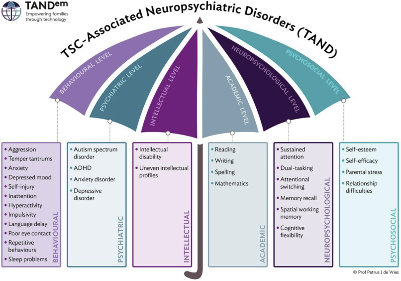
Neurodevelopmental disorder is an important feature in tuberous sclerosis. Images 3 and 4 (Figures 3 and 4) show brain abnormalities that may indicate neurological developmental issues.
Figure 9. Image 3 shows alterations in the brain associated with the neurodevelopmental disorder in tuberous sclerosis, with evidence of lesions in the central nervous system, such as cysts and malformations of neuronal development.Figure 10. Image 4 illustrates neuroradiological changes in the brain of patients with tuberous sclerosis, highlighting the presence of lesions in the central nervous system, such as white matter tumors and lesions in the striate ganglion.
Neuroradiological Manifestations
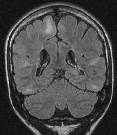
Neuroradiological manifestations are critical for the diagnosis and monitoring of tuberous sclerosis. Image 5 (Figure 5) shows the typical radiological features of the disease.
Figure 11. Image 5 shows the typical radiological features of tuberous sclerosis on magnetic resonance images, with multiple lesions in the central nervous system, including tumors in the white matter and lesions in the ventricular system.Figure 5: Neuroradiological manifestations of tuberous sclerosis.
Systemic Complications Onset Timeline
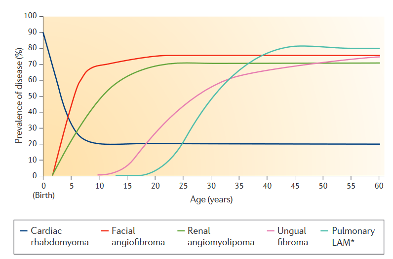
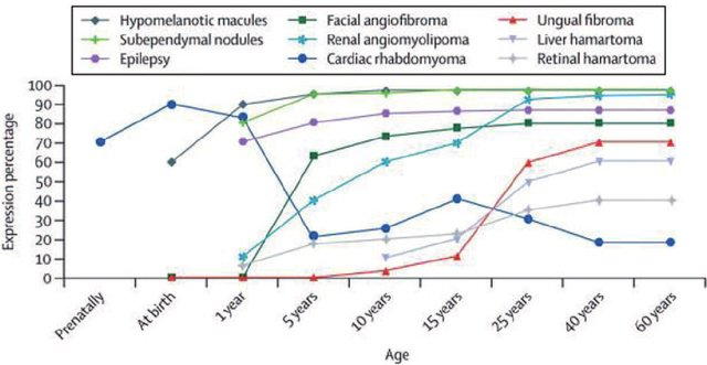
Tuberous sclerosis can present with a range of systemic complications over time. Figure 6 illustrates the chronological appearance of these complications.
Figure 12. Image 6 illustrates the chronology of appearance of systemic complications in tuberous sclerosis, showing how damage to organs such as the kidney, heart and lung can develop over time after the onset of the disease.Figure 6: Timeline of systemic complications in tuberous sclerosis.
Regarding tuberous sclerosis, radiology can be a valuable resource for identifying lesions. According to Radiopaedia.org, the radiological characteristics include lesions in the brain, skin, and other body structures consistent with tuberous sclerosis. Interpreting these images requires detailed clinical understanding of the disease's course and associated radiological patterns.