1. Introduction and Relevance of Neuroimaging
The Brain Magnetic Resonance (MRI) constitutes the first-line complementary test after the anamnesis and detailed neurological examination in all children with suspected Infantile Cerebral Palsy (CPI). It is estimated that MRI reveals pathogenic abnormalities in more than 80% of cases, orienting on the temporal moment and the nature of the cerebral noxa.
2. SCPE Classification according to Neuroimaging Patterns
The network of Surveillance of Cerebral Palsy in Europe (SCPE) Stratifies neuroimaging findings into 5 essential categories:
- Predominant white matter lesions (approx. 50%): Periventricular leukomalacia, typically secondary to perinatal ischemic or infectious events in premature infants.
- Lesions of cortical gray matter and basal ganglia (approx. 20%): Hypoxic-ischemic lesions at term or acute perinatal encephalopathies.
- Brain development malformations (approx. 11%): Migration disorders, polymicrogyria, schicencephaly or lissencephaly.
- Normal Magnetic Resonance (approx. 11%): Absence of obvious acquired structural damage.
- Miscellaneous/Nonspecific: Other anatomical alterations not categorizable in the previous groups.
3. When to Indicate Genetic Studies in PCI?
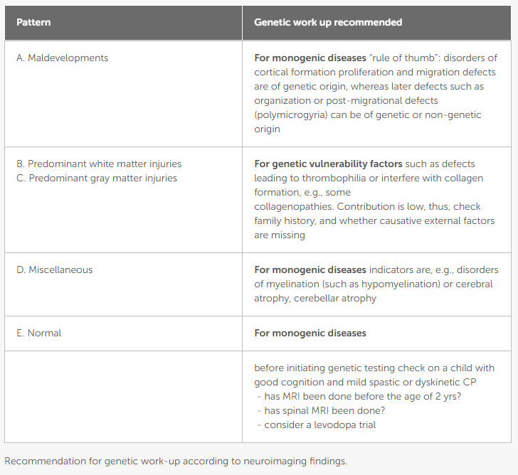
The diagnostic performance of the genetic study (chromosomal microarrays, massive sequencing panels or whole exome) varies drastically depending on the neuroimaging pattern:
- Priority Indication and High Monogenic Suspicion: In patients with normal MRI or with brain development malformations, the probability of an underlying monogenic etiology is very high. In these cases, genetic study is formally indicated.
- Genetic Vulnerability in Acquired Injuries: In cases with acquired white or gray matter lesions without a clear history of asphyxia or perinatal risk factors, genetic predisposition that increases cellular vulnerability to minimal noxas should be considered.
4. Practical Recommendations for Consultation
In a patient with symptoms of cerebral palsy without a clear perinatal history or with an atypical or normal neuroimaging, one should avoid assuming an undocumented cause of injury and start the diagnostic genetic algorithm early.
Bibliography and References
19955111
{19955111:NMMWTEGD}
1
vancouver
50
default
8025
https://neuropediatoolkit.org/wp-content/plugins/zotpress/
%7B%22status%22%3A%22success%22%2C%22updateneeded%22%3Afalse%2C%22instance%22%3Afalse%2C%22meta%22%3A%7B%22request_last%22%3A0%2C%22request_next%22%3A0%2C%22used_cache%22%3Atrue%7D%2C%22data%22%3A%5B%7B%22key%22%3A%22NMMWTEGD%22%2C%22library%22%3A%7B%22id%22%3A19955111%7D%2C%22meta%22%3A%7B%22creatorSummary%22%3A%22Horber%20et%20al.%22%2C%22parsedDate%22%3A%222021-02-09%22%2C%22numChildren%22%3A1%7D%2C%22bib%22%3A%22%26lt%3Bdiv%20class%3D%26quot%3Bcsl-bib-body%26quot%3B%20style%3D%26quot%3Bline-height%3A%201.35%3B%20%26quot%3B%26gt%3B%5Cn%20%20%26lt%3Bdiv%20class%3D%26quot%3Bcsl-entry%26quot%3B%20style%3D%26quot%3Bclear%3A%20left%3B%20%26quot%3B%26gt%3B%5Cn%20%20%20%20%26lt%3Bdiv%20class%3D%26quot%3Bcsl-left-margin%26quot%3B%20style%3D%26quot%3Bfloat%3A%20left%3B%20padding-right%3A%200.5em%3B%20text-align%3A%20right%3B%20width%3A%201em%3B%26quot%3B%26gt%3B1.%26lt%3B%5C%2Fdiv%26gt%3B%26lt%3Bdiv%20class%3D%26quot%3Bcsl-right-inline%26quot%3B%20style%3D%26quot%3Bmargin%3A%200%20.4em%200%201.5em%3B%26quot%3B%26gt%3BHorber%20V%2C%20Grasshoff%20U%2C%20Sellier%20E%2C%20Arnaud%20C%2C%20Kr%26%23xE4%3Bgeloh-Mann%20I%2C%20Himmelmann%20K.%20The%20Role%20of%20Neuroimaging%20and%20Genetic%20Analysis%20in%20the%20Diagnosis%20of%20Children%20With%20Cerebral%20Palsy.%20Front%20Neurol%20%5BInternet%5D.%202021%20Feb%209%20%5Bcited%202026%20Jan%204%5D%3B11.%20Available%20from%3A%20%26lt%3Ba%20class%3D%26%23039%3Bzp-ItemURL%26%23039%3B%20href%3D%26%23039%3Bhttps%3A%5C%2F%5C%2Fwww.frontiersin.org%5C%2Fjournals%5C%2Fneurology%5C%2Farticles%5C%2F10.3389%5C%2Ffneur.2020.628075%5C%2Ffull%26%23039%3B%26gt%3Bhttps%3A%5C%2F%5C%2Fwww.frontiersin.org%5C%2Fjournals%5C%2Fneurology%5C%2Farticles%5C%2F10.3389%5C%2Ffneur.2020.628075%5C%2Ffull%26lt%3B%5C%2Fa%26gt%3B%26lt%3B%5C%2Fdiv%26gt%3B%5Cn%20%20%26lt%3B%5C%2Fdiv%26gt%3B%5Cn%26lt%3B%5C%2Fdiv%26gt%3B%22%2C%22data%22%3A%7B%22itemType%22%3A%22journalArticle%22%2C%22title%22%3A%22The%20Role%20of%20Neuroimaging%20and%20Genetic%20Analysis%20in%20the%20Diagnosis%20of%20Children%20With%20Cerebral%20Palsy%22%2C%22creators%22%3A%5B%7B%22creatorType%22%3A%22author%22%2C%22firstName%22%3A%22Veronka%22%2C%22lastName%22%3A%22Horber%22%7D%2C%7B%22creatorType%22%3A%22author%22%2C%22firstName%22%3A%22Ute%22%2C%22lastName%22%3A%22Grasshoff%22%7D%2C%7B%22creatorType%22%3A%22author%22%2C%22firstName%22%3A%22Elodie%22%2C%22lastName%22%3A%22Sellier%22%7D%2C%7B%22creatorType%22%3A%22author%22%2C%22firstName%22%3A%22Catherine%22%2C%22lastName%22%3A%22Arnaud%22%7D%2C%7B%22creatorType%22%3A%22author%22%2C%22firstName%22%3A%22Ingeborg%22%2C%22lastName%22%3A%22Kr%5Cu00e4geloh-Mann%22%7D%2C%7B%22creatorType%22%3A%22author%22%2C%22firstName%22%3A%22Kate%22%2C%22lastName%22%3A%22Himmelmann%22%7D%5D%2C%22abstractNote%22%3A%22Cerebral%20magnetic%20resonance%20imaging%20%28MRI%29%20is%20considered%20an%20important%20tool%20in%20the%20assessment%20of%20a%20child%20with%20cerebral%20palsy%20%28CP%29%2C%20as%20it%20is%20abnormal%20in%20more%20than%2080%25%20of%20children%20with%20CP%2C%20disclosing%20the%20pathogenic%20pattern%20responsible%20for%20the%20neurological%20condition.%20MRI%2C%20therefore%2C%20is%20recommended%20as%20the%20first%20diagnostic%20step%20after%20medical%20history%20taking%20and%20neurological%20examination.With%20the%20advances%20in%20genetic%20diagnostics%2C%20the%20genetic%20contribution%20to%20CP%20is%20increasingly%20discussed%20and%20the%20question%20arises%20about%20the%20role%20of%20genetic%20testing%20in%20the%20diagnosis%20of%20cerebral%20palsy.%20The%20paper%20gives%20an%20overview%20on%20genetic%20findings%20reported%20in%20CP%2C%20which%20are%20discussed%20with%20respect%20to%20the%20underlying%20brain%20pathology%20according%20to%20neuroimaging%20findings.Surveillance%20of%20Cerebral%20Palsy%20in%20Europe%20%28SCPE%29%20classifies%20neuroimaging%20findings%20in%20CP%20into%20five%20categories%2C%20which%20help%20to%20stratify%20decisions%20concerning%20genetic%20testing.%20Predominant%20white%20and%20grey%20matter%20injuries%20are%20by%20far%20predominant%20%28accounting%20for%20around%2050%25%20and%2020%25%20of%20findings%29.%20They%20are%20considered%20to%20be%20acquired.%20Here%2C%20predisposing%20genetic%20factors%20may%20play%20a%20role%20to%20increase%20vulnerability%20%28and%20should%20especially%20be%20considered%2C%20when%20family%20history%20is%20positive%20and%5C%2For%20causative%20external%20factors%20are%20missing%29.%20In%20maldevelopments%20and%20normal%20findings%20%28around%2011%25%20each%29%2C%20monogenic%20causes%20are%20more%20likely%20and%20thus%2C%20genetic%20testing%20is%20clearly%20recommended.%20In%20the%20miscellaneous%20category%2C%20the%20precise%20nature%20of%20the%20MRI%20finding%20has%20to%20be%20considered%20as%20it%20could%20indicate%20a%20genetic%20origin.%22%2C%22date%22%3A%222021-02-09%22%2C%22section%22%3A%22%22%2C%22partNumber%22%3A%22%22%2C%22partTitle%22%3A%22%22%2C%22DOI%22%3A%2210.3389%5C%2Ffneur.2020.628075%22%2C%22citationKey%22%3A%22%22%2C%22url%22%3A%22https%3A%5C%2F%5C%2Fwww.frontiersin.org%5C%2Fjournals%5C%2Fneurology%5C%2Farticles%5C%2F10.3389%5C%2Ffneur.2020.628075%5C%2Ffull%22%2C%22PMID%22%3A%22%22%2C%22PMCID%22%3A%22%22%2C%22ISSN%22%3A%221664-2295%22%2C%22language%22%3A%22English%22%2C%22collections%22%3A%5B%22D3GNDRTB%22%5D%2C%22dateModified%22%3A%222026-07-15T22%3A38%3A27Z%22%7D%7D%5D%7D