
1. ニューロイメージングの概要と関連性
の 脳磁気共鳴 (MRI) 疑いのあるすべての小児の既往歴と詳細な神経学的検査後の第一選択の補完検査を構成します。 乳児脳性麻痺 (CPI)。 MRI は、以下のものの病原性異常を明らかにすると推定されています。 80%のケース、時間的瞬間と大脳ノクサの性質を重視しています。
2. 神経画像パターンによる SCPE の分類
のネットワーク ヨーロッパにおける脳性麻痺の監視 (SCPE) 神経画像所見を 5 つの重要なカテゴリに階層化します。
- 主な白質病変 (約 50%): 脳室周囲白質軟化症。通常、未熟児における周産期虚血性イベントまたは感染症イベントに続発します。
- 皮質灰白質および大脳基底核の病変 (約 20%): 正期産または急性周産期脳症における低酸素性虚血性病変。
- 脳の発達奇形 (約 11%): 移動障害、多小脳症、大脳症または滑脳症。
- 通常の磁気共鳴 (約 11%): 明らかな後天的な構造的損傷がないこと。
- その他/不特定: 前のグループに分類できないその他の解剖学的変化。
3. PCI で遺伝的研究を示すのはどのような場合ですか?
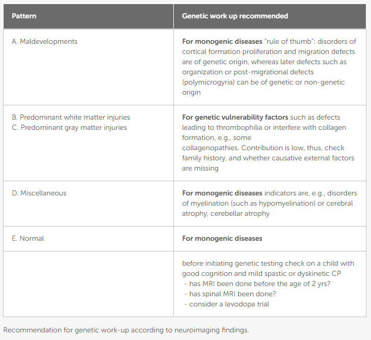
遺伝子研究 (染色体マイクロアレイ、大規模配列決定パネル、または全エクソーム) の診断パフォーマンスは、神経画像パターンに応じて大幅に異なります。
- 優先適応症と単一遺伝子性の高い疑い: 患者の場合 通常のMRI または一緒に 脳の発達奇形、根底にある単一遺伝子の病因の可能性は非常に高いです。このような場合、遺伝子研究が正式に指示されます。
- 後天性損傷における遺伝的脆弱性: 窒息や周産期の危険因子の明らかな病歴がない後天性の白質または灰白質病変の場合、最小限のノクサスに対する細胞の脆弱性を高める遺伝的素因を考慮する必要があります。
4. 相談に関する実際的な推奨事項
明らかな周産期病歴のない脳性麻痺の症状がある患者、または神経画像検査が非定型または正常である患者の場合は、文書化されていない傷害の原因を仮定することを避け、診断の遺伝的アルゴリズムを早期に開始する必要があります。
参考文献と参考文献
19955111
{19955111:NMMWTEGD}
1
バンクーバー
50
デフォルト
8025
https://neuropediatoolkit.org/wp-content/plugins/zotpress/
%7B%22status%22%3A%22success%22%2C%22updateneeded%22%3Afalse%2C%22instance%22%3Afalse%2C%22meta%22%3A%7B%22request_last%22%3A0%2C%22request_next%22%3A0%2C%22used_cache%22%3Atrue%7D%2C%22data%22%3A%5B%7B%22key%22%3A%22NMMWTEGD%22%2C%22library%22%3A%7B%22id%22%3A19955111%7D%2C%22meta%22%3A%7B%22creatorSummary%22%3A%22Horber%20et%20al.%22%2C%22parsedDate%22%3A%222021-02-09%22%2C%22numChildren%22%3A1%7D%2C%22bib%22%3A%22%26lt%3Bdiv%20class%3D%26quot%3Bcsl-bib-body%26quot%3B%20style%3D%26quot%3Bline-height%3A%201.35%3B%20%26quot%3B%26gt%3B%5Cn%20%20%26lt%3Bdiv%20class%3D%26quot%3Bcsl-entry%26quot%3B%20style%3D%26quot%3Bclear%3A%20left%3B%20%26quot%3B%26gt%3B%5Cn%20%20%20%20%26lt%3Bdiv%20class%3D%26quot%3Bcsl-left-margin%26quot%3B%20style%3D%26quot%3Bfloat%3A%20left%3B%20padding-right%3A%200.5em%3B%20text-align%3A%20right%3B%20width%3A%201em%3B%26quot%3B%26gt%3B1.%26lt%3B%5C%2Fdiv%26gt%3B%26lt%3Bdiv%20class%3D%26quot%3Bcsl-right-inline%26quot%3B%20style%3D%26quot%3Bmargin%3A%200%20.4em%200%201.5em%3B%26quot%3B%26gt%3BHorber%20V%2C%20Grasshoff%20U%2C%20Sellier%20E%2C%20Arnaud%20C%2C%20Kr%26%23xE4%3Bgeloh-Mann%20I%2C%20Himmelmann%20K.%20The%20Role%20of%20Neuroimaging%20and%20Genetic%20Analysis%20in%20the%20Diagnosis%20of%20Children%20With%20Cerebral%20Palsy.%20Front%20Neurol%20%5BInternet%5D.%202021%20Feb%209%20%5Bcited%202026%20Jan%204%5D%3B11.%20Available%20from%3A%20%26lt%3Ba%20class%3D%26%23039%3Bzp-ItemURL%26%23039%3B%20href%3D%26%23039%3Bhttps%3A%5C%2F%5C%2Fwww.frontiersin.org%5C%2Fjournals%5C%2Fneurology%5C%2Farticles%5C%2F10.3389%5C%2Ffneur.2020.628075%5C%2Ffull%26%23039%3B%26gt%3Bhttps%3A%5C%2F%5C%2Fwww.frontiersin.org%5C%2Fjournals%5C%2Fneurology%5C%2Farticles%5C%2F10.3389%5C%2Ffneur.2020.628075%5C%2Ffull%26lt%3B%5C%2Fa%26gt%3B%26lt%3B%5C%2Fdiv%26gt%3B%5Cn%20%20%26lt%3B%5C%2Fdiv%26gt%3B%5Cn%26lt%3B%5C%2Fdiv%26gt%3B%22%2C%22data%22%3A%7B%22itemType%22%3A%22journalArticle%22%2C%22title%22%3A%22The%20Role%20of%20Neuroimaging%20and%20Genetic%20Analysis%20in%20the%20Diagnosis%20of%20Children%20With%20Cerebral%20Palsy%22%2C%22creators%22%3A%5B%7B%22creatorType%22%3A%22author%22%2C%22firstName%22%3A%22Veronka%22%2C%22lastName%22%3A%22Horber%22%7D%2C%7B%22creatorType%22%3A%22author%22%2C%22firstName%22%3A%22Ute%22%2C%22lastName%22%3A%22Grasshoff%22%7D%2C%7B%22creatorType%22%3A%22author%22%2C%22firstName%22%3A%22Elodie%22%2C%22lastName%22%3A%22Sellier%22%7D%2C%7B%22creatorType%22%3A%22author%22%2C%22firstName%22%3A%22Catherine%22%2C%22lastName%22%3A%22Arnaud%22%7D%2C%7B%22creatorType%22%3A%22author%22%2C%22firstName%22%3A%22Ingeborg%22%2C%22lastName%22%3A%22Kr%5Cu00e4geloh-Mann%22%7D%2C%7B%22creatorType%22%3A%22author%22%2C%22firstName%22%3A%22Kate%22%2C%22lastName%22%3A%22Himmelmann%22%7D%5D%2C%22abstractNote%22%3A%22Cerebral%20magnetic%20resonance%20imaging%20%28MRI%29%20is%20considered%20an%20important%20tool%20in%20the%20assessment%20of%20a%20child%20with%20cerebral%20palsy%20%28CP%29%2C%20as%20it%20is%20abnormal%20in%20more%20than%2080%25%20of%20children%20with%20CP%2C%20disclosing%20the%20pathogenic%20pattern%20responsible%20for%20the%20neurological%20condition.%20MRI%2C%20therefore%2C%20is%20recommended%20as%20the%20first%20diagnostic%20step%20after%20medical%20history%20taking%20and%20neurological%20examination.With%20the%20advances%20in%20genetic%20diagnostics%2C%20the%20genetic%20contribution%20to%20CP%20is%20increasingly%20discussed%20and%20the%20question%20arises%20about%20the%20role%20of%20genetic%20testing%20in%20the%20diagnosis%20of%20cerebral%20palsy.%20The%20paper%20gives%20an%20overview%20on%20genetic%20findings%20reported%20in%20CP%2C%20which%20are%20discussed%20with%20respect%20to%20the%20underlying%20brain%20pathology%20according%20to%20neuroimaging%20findings.Surveillance%20of%20Cerebral%20Palsy%20in%20Europe%20%28SCPE%29%20classifies%20neuroimaging%20findings%20in%20CP%20into%20five%20categories%2C%20which%20help%20to%20stratify%20decisions%20concerning%20genetic%20testing.%20Predominant%20white%20and%20grey%20matter%20injuries%20are%20by%20far%20predominant%20%28accounting%20for%20around%2050%25%20and%2020%25%20of%20findings%29.%20They%20are%20considered%20to%20be%20acquired.%20Here%2C%20predisposing%20genetic%20factors%20may%20play%20a%20role%20to%20increase%20vulnerability%20%28and%20should%20especially%20be%20considered%2C%20when%20family%20history%20is%20positive%20and%5C%2For%20causative%20external%20factors%20are%20missing%29.%20In%20maldevelopments%20and%20normal%20findings%20%28around%2011%25%20each%29%2C%20monogenic%20causes%20are%20more%20likely%20and%20thus%2C%20genetic%20testing%20is%20clearly%20recommended.%20In%20the%20miscellaneous%20category%2C%20the%20precise%20nature%20of%20the%20MRI%20finding%20has%20to%20be%20considered%20as%20it%20could%20indicate%20a%20genetic%20origin.%22%2C%22date%22%3A%222021-02-09%22%2C%22section%22%3A%22%22%2C%22partNumber%22%3A%22%22%2C%22partTitle%22%3A%22%22%2C%22DOI%22%3A%2210.3389%5C%2Ffneur.2020.628075%22%2C%22citationKey%22%3A%22%22%2C%22url%22%3A%22https%3A%5C%2F%5C%2Fwww.frontiersin.org%5C%2Fjournals%5C%2Fneurology%5C%2Farticles%5C%2F10.3389%5C%2Ffneur.2020.628075%5C%2Ffull%22%2C%22PMID%22%3A%22%22%2C%22PMCID%22%3A%22%22%2C%22ISSN%22%3A%221664-2295%22%2C%22language%22%3A%22English%22%2C%22collections%22%3A%5B%22D3GNDRTB%22%5D%2C%22dateModified%22%3A%222026-07-15T22%3A38%3A27Z%22%7D%7D%5D%7D
1.
Horber V、Grasshoff U、Sellier E、Arnaud C、Krägeloh-Mann I、Himmelmann K. 脳性麻痺の小児の診断における神経画像診断と遺伝子分析の役割。フロントニューロール [インターネット]。 2021 2 9 [2026 1 4 引用];11.以下から入手可能: https://www.frontiersin.org/journals/neurology/articles/10.3389/fneur.2020.628075/full