
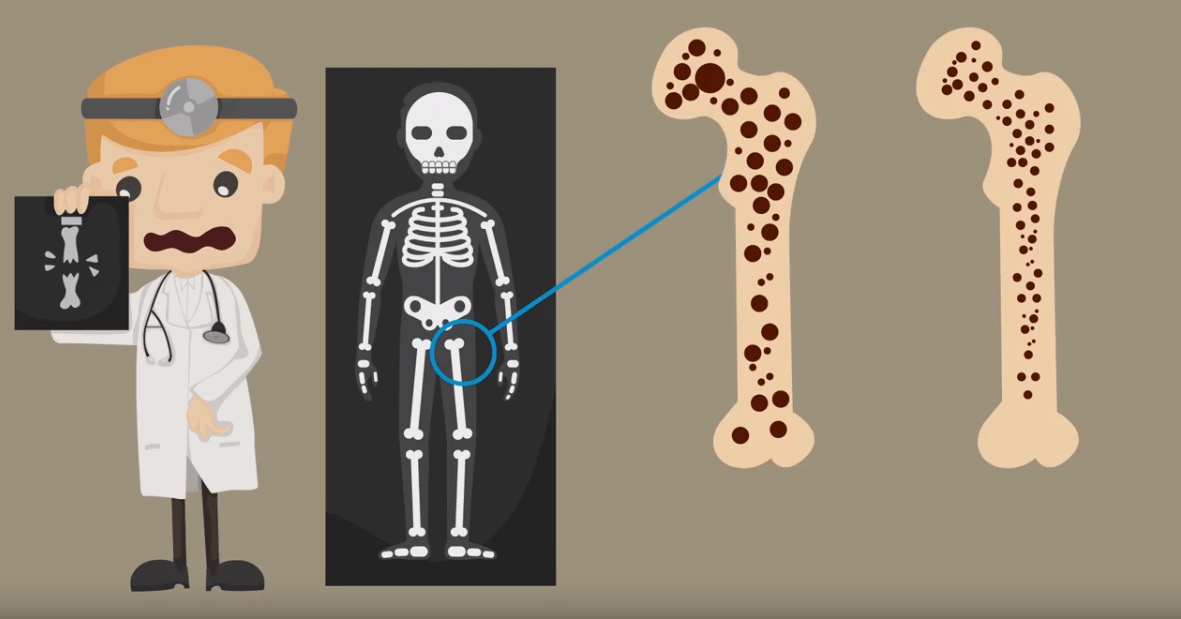
The childhood osteoporosis It is defined, in clinical practice, by the presence of fragility fractures.
The International Society of Clinical Densitometry (ISCD), in its 2019 consensus, establishes two alternative conditions for the diagnosis [1,2]:
- Presence of one or more vertebral fractures in the absence of local disease or high-energy trauma.
- BMD or CMO Z-score ≤ −2 (adjusted to size if it is < 3rd percentile), associated to a history of clinically significant fractures, meaning:
- Two or more long bone fractures before the age of 10, o
- Three or more long bone fractures before age 19.
Important: a Z-score > −2 does not exclude bone fragility or predisposition to fractures, especially in patients with pathologies that favor secondary osteoporosis [1,2].
Risk factors for secondary osteoporosis
- Immobility
- Pubertal delay
- Little sun exposure
- Nutritional deficiency
- Sustained inflammatory activity
- Osteotoxic drugs (corticosteroids, antiepileptics) [1]
Imaging technique according to age
| Age | Recommended DXA location |
|---|---|
| Infants and preschoolers (< 5 years) | lumbar spine |
| ≥ 3 years | Whole body (excluding head) |
Calculation and reference resources:
- Z-score calculator (CHOP): https://zscore.research.chop.edu/calcpedbonedens.php
- Food Composition Database (BEDCA): https://www.bedca.net/bdpub/index.php
Vitamin D: dosage and supplementation
Recommended daily dose in healthy children [1,6]:
- < 1 year: 400 IU/day
- > 1 year: 600 IU/day
The American Academy of Pediatrics recommends supplementing with vitamin D in [1,6]:
- Every newborn, from the first days of life to one year.
- Children 1-2 years old who must drink milk or skimmed milk products.
- Older children who drink less than 1 L/day of milk not fortified with vitamin D.
- Adolescents who do not reach 400 IU/day.
- Children at special risk of deficiency (malabsorption, antiepileptic medication): higher doses based on serum 25-OH-vitamin D levels, with periodic monitoring.
Note: There is currently no daily dose of calcium and vitamin D specifically established for children with chronic pathology. In these patients, with low BMD for chronological age, it is recommended to supplement at the reference doses of the healthy child and adjust according to iPTH, calciuria and vitamin D levels [1].
Associated general measures:
- Diet rich in fruit and vegetables.
- In oral or inhaled corticosteroid therapy: supplement with calcium and vitamin D if the prednisone dose is ≥ 5 mg/day [1,9].
Bisphosphonates in childhood osteoporosis
General indication
The most recent consensus on the use of bisphosphonates in children and adolescents recommends considering them whenever osteoporosis criteria are met, with the aim of prevent new fragility fractures [10,12].
Use in patients without osteoporosis but with low BMD
They can be assessed in patients without osteoporosis criteria but with low BMD at the onset of puberty (Tanner 2), in two situations [10]:
- If risk factors persist and the Z-score < −2.5 with a downward trend in two determinations separated by at least one year.
- In the absence of risk factors, if the Z-score < −3 with a downward trend in two determinations separated by at least one year.
Tolerance and side effects
In general they are well tolerated. With the intravenous bisphosphonates may appear [11]:
- Flu-like syndrome: fever, malaise, nausea, diarrhea and generalized myalgia, typically between the 1st and 3rd day of treatment; It lasts a few days and rarely recurs in successive doses. Responds to paracetamol.
- Hypocalcemia, usually asymptomatic; occasionally arrhythmias, laryngospasm or tetany.
- Hypophosphoremia and hypomagnesemia asymptomatic.
- Others, described in adults but exceptional in pediatrics: uveitis, thrombopenia, oral or esophageal ulcers, osteonecrosis of the jaw [11].
Measures to reduce the risk of adverse effects
- Determine ionic calcium 48 hours after the infusion.
- Ensure adequate levels of vitamin D and calcium before to start treatment.
- Dental check-up before the start and every 6 months during therapy.
- Suspend treatment 3 to 4 months before any elective osteotomy, and restart it only when the callus is formed [14].
- Kidney ultrasound to detect nephrocalcinosis in relation to vitamin D supplementation.
- Monitor calcium levels of newborns of mothers treated with bisphosphonates before or during pregnancy [15].
Administration schedule (pamidronate/zoledronate)
- Before each infusion, check: vitamin D ≥ 30 ng/dL, normal calcium and kidney function.
- On the first infusion, administer half the dose.
- Manage paracetamol before the infusion to reduce side effects (fever, pain).
- Administer (or increase) supplements calcium the week before and the week after treatment [11].
Follow-up
DXA
Frequency depending on the treatment received:
- Prolonged corticosteroid therapy: annually or biannually for the duration of the treatment, and even every 6 months if necessary. The first evaluation is recommended 3 months after the start of corticosteroid therapy [4].
- Chronic diseases without corticosteroids: there is no consensus; individualize according to:
- underlying disease
- Bone quality at initial evaluation
- Nutritional status
- Life habits
- Occurrence of fractures
Other explorations
- spine x-ray: annual, if low bone mass or risk factor persists.
- Analytics: annual.
- Kidney ultrasound: if hypercalciuria, to rule out nephrocalcinosis.
Literature
- Galindo Zavala R, Núñez Cuadros E, Díaz-Cordovés Rego G. States of low bone mineralization. Childhood osteoporosis. Protoc Diagn Ter Pediatr. 2020;2:335-348. Available at: https://www.aeped.es/protocolos/
- The International Society for Clinical Densitometry. 2019 ISCD Official Positions – Pediatric. Available at: https://www.iscd.org/official-positions/2019-iscd-official-positions-pediatric/
- Marrani E, Giani T, Simonini G, Cimaz R. Pediatric Osteoporosis: Diagnosis and Treatment Considerations. Drugs. 2017;77:679-95.
- Ward LM, Konji VN, Ma J. The management of osteoporosis in children. Osteoporos Int. 2016;27:2147-79.
- Di Iorgi N, Maruca K, Patti G, Mora S. Update on bone density measurements and their interpretation in children and adolescents. Best Pract Res Clin Endocrinol Metab. 2018;32:477-98.
- Wagner CL, Greer FR. Prevention of ricketts and vitamin D deficiency in infants, children, and adolescents. Pediatrics. 2008;122:1142-52.
- Hillman LS, Cassidy JT, Chanetsa F, et al. Percent true calcium absorption, mineral metabolism, and bone mass in children with arthritis: effect of supplementation with vitamin D3 and calcium. Rheum Arthritis. 2008;58:3255-63.
- Galindo Zavala R, Bou Torrent R, Magallares López B, et al. Expert panel consensus recommendations for diagnosis and treatment of secondary osteoporosis in children. Pediatr Rheumatol Online J. 2020;18(1):20.
- Bachrach LK, Ward LM. Clinical review 1: Bisphosphonate use in childhood osteoporosis. J Clin Endocrinol Metab. 2009;94:400-9.
- Simm PJ, Biggin A, Zacharin MR, et al. Consensus guidelines on the use of bisphosphonate therapy in children and adolescents. J Paediatr Child Health. 2018;54:223-33.
- Munns CF, Rauch F, Zeitlin L, Fassier F, Glorieux FH. Delayed osteotomy but not fracture healing in pediatric osteogenesis imperfecta patients receiving pamidronate. J Bone Miner Res. 2004;19:1779-86.
- Djokanovic N, Klieger-Grossmann C, Koren G. Does treatment with bisphosphonates endanger the human pregnancy? J Obstet Gynaecol Can. 2008;30:1146-8.
Additional resources cited in the text:
- Pediatric Z-score Calculator (CHOP): https://zscore.research.chop.edu/calcpedbonedens.php
- Spanish food composition database (BEDCA): https://www.bedca.net/bdpub/index.php